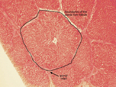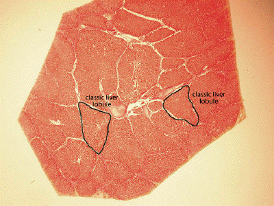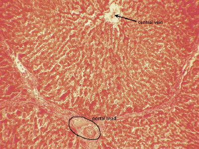
Slide #DMS 144 [Liver, Pig, trichrome stain (collagen stains blue)]. Pig liver has abundant interlobular fibrous connective tissue. Because of this, the "classic liver lobule" is well demarcated. In the centers of most lobules can be seen a central vein. Situated in the interlobular connective tissue, mostly at angles, are portal triads, where branches of the portal vein, hepatic artery, and bile duct and often a lymphatic vessel can be found. Unfortunately, this particular specimen is not too well preserved (Probably it sat, unfixed, in a meat market for several days) so that some of the structures (particularly veins) in some triads are torn or otherwise distorted. However, if you look around you can find intact triads.

Begin with this very low power overview of a section through the pig liver. Unlike the human liver, an abundance of interlobular connective tissue is seen in the pig liver which clearly demarcates the classic liver lobule.

At low power of this trichrome-stained pig liver, the boundaries of the classic liver lobule are clearly demarcated. At the center of the classic liver lobule, one finds the central vein. Around the periphery of the lobule, one may find elements of the portal triad.

A medium power view of the pig liver shows a portion of a classic liver lobule with the central vein at the top of the image, and components of a portal triad at the bottom. A more detailed inspection of these elements will be possible in subsequent slides in which the tissue preservation is superior.