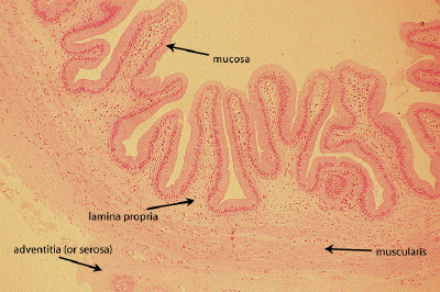
Slide #DMS 150 [Gallbladder, Human, PTS]. Observe that the mucosa consists of a simple columnar epithelium and a lamina propria. The lamina propria here was studied earlier as a well preserved example of loose (areolar) connective tissue. There is no muscularis mucosa or submucosa. Note that the muscularis is in irregular bundles that does not show the inner circular and outer longitudinal arrangement found in the gut. External to the muscularis is some moderately dense connective tissue. Using higher power, return to the mucosa. Note that it is thrown up into folds. The columnar epithelial cells are quite tall. Goblet cells are not normally found in the epithelium. A section of gall bladder should not be confused with sections taken elsewhere in the gastro-intestinal tract. The gall bladder has no villi, no crypts, no muscularis mucosa and no goblet cells in its epithelium. There is an external serosa with a mesothelial surface. What is the function of the gall bladder?
This image provides a low power view of another section of gall bladder. At this magnification, one might again confuse this organ with small intestine because of the villous-like mucosal folds. Again, identify the various layers forming the thickness of the gall bladder: mucosa, lamina propria, muscularis, adventitia (or serosa).
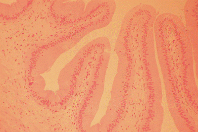
At medium power, one can appreciate the homogeneity of the surface epithelium, a simple columnar epithelium devoid of goblet cells.
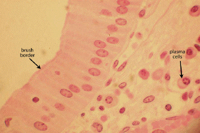
At high power, the apical surface of the gall bladder epithelial cells may be seen to exhibit a short brush border. Note the distinctive plasma cells within the underlying lamina propria