Slide #DMS 104 [Marrow smear, Wright Stain].
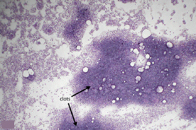
This is a low power view of a marrow smear, the preferred preparation for examining the morphology of hematopoietic cells. The cells streaming away from the small clots are often of good morphological quality and will be examined in subsequent images.
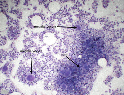
A medium power view of a marrow smear shows a small clot surrounded by individually-distinguishable marrow cells, the details of which are really best appreciated with 100x oil immersion optics. At this power, only the large, multilobulated megakaryocytes can be unequivocally identified.
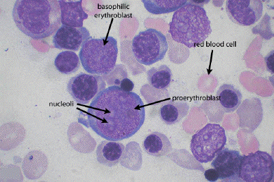
This oil immersion image shows the characteristic features of the early stages of erythroid development. Using mature red blood cells as a yardstick, note the size of their developing precursors, beginning with the proerythroblast. This large cell is characterized by a large, round nucleus containing one or more nucleoli (lightly stained). The cytoplasm of this cell may show an intense royal blue basophilia. As development proceeds, these cells will become smaller, the nucleoli will disappear, but the royal blue basophilic cytoplasm will persist into the basophilic erythroblast stage. Remember that hematopoiesis represents a developmental continuum, and that many of the cells seen will be somewhere in between the classically-defined stages.
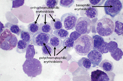
Compare the size, nuclear morphology and cytoplasmic staining of the basophilic erythroblast (baso) with subsequent stages of development, the polychromatophilic erythroblast (poly) and then the orthochromatophilic erythroblast (ortho). The polys are characterized by a grayish-blue cytoplasm, and a round nucleus with a 'checkerboard' chromatin pattern. With further development, these cells continue to shrink, the nucleus continues to condense, and the increased production of hemoglobin gives the cytoplasm of the orthochromatophilic erythroblasts a faintly pinkish hue (but compare to mature RBC).
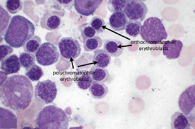
In this image, note the changes in size, degree of nuclear condensation, and cytoplasmic coloration as one progresses from polychromatophilic erythroblasts to the orthochromatophilic erythroblasts.
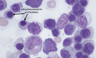
In this image, note the two orthochromatophilic erythroblasts, with rather eosinophilic cytoplasm (still not yet of the mature hue, however) and dark, condensed nuclei. After the extrusion of their nuclei, these cells will then be called reticulocytes prior to their final maturation into erythrocytes.

This image illustrates the feature of two promyelocytes, one of the early stages in the development of the granulocytes. These large cells are characterized by multiple nucleoli, and a lightly basophilic cytoplasm in which one may find rather large, azurophilic granules (aka primary granules or lysosomes). Prior to the appearance of the secondary or specific granules, it is not morphologically clear which of the granulocytes these promyelocytes will become.
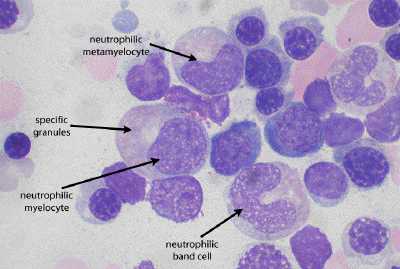
With the appearance of secondary or specific granules at the myelocyte stage, one may now identify the particular granulocyte lineage. Here, one sees a neutrophilic myelocyte, characterized by a round, flattened or just slightly-indented nucleus lacking nucleoli. Most notably, an abundance of specific granules is apparent and gives the cytoplasm a somewhat salmon-colored hue. The primary or azurophilic granules are usually not so apparent at this stage. Following the myelocyte stage, the nucleus begins to indent more noticeably yielding a neutrophilic metamyelocyte. As the nucleus continues to thin out, this cell will progress into the neutrophilic band cell, an early stage example of which is seen here.
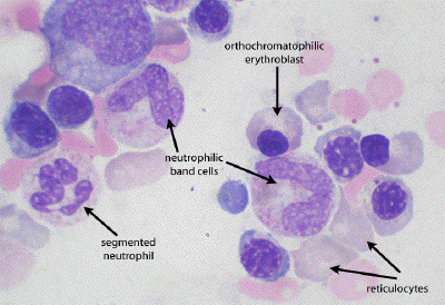
This image shows two classic neutrophilic band cells, the nuclei of which may begin to fold on themselves, yielding rather odd-looking nuclei. Continued maturation of the band cell will yield a mature, segmented neutrophil characterized by a multi-lobed nucleus with the lobes connected by thin strands of chromatin. In this image, note again an orthochromatophilic erythroblast, and a couple of reticulocytes (compare size and hue to the mature RBCs).
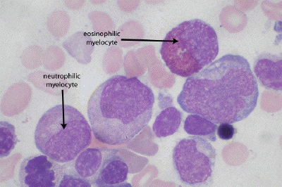
As an example of one of the other granulocytes, compare the morphology of a neutrophilic myelocyte with an eosinophilic myelocyte. The secondary or specific granules of the eosinophil lineage are noticeably larger than those found in neutrophils which are barely resolvable and simply give the neutrophil cytoplasm a salmon-colored, slightly grainy texture.
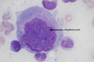
Developing megakaryocytes are impossible to miss given their huge size relative to the other hematopoietic cells. As this cells develops, its nucleus will become increasingly lobulated as polyploidy develops.
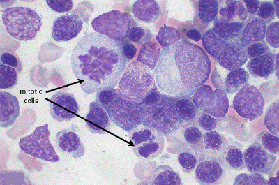
In addition to developing cells corresponding to all the classically-defined developmental stages, appreciate that mitosis is in integral part of this process. Cells in the midst of mitosis, identified by their condensed chromosomes, are not an uncommon sight in marrow smears.