Slide #DMS 075 [Red bone marrow]. This is a section of rib from a young rabbit. Compare the appearance of the red marrow elements to those seen in Slide #103. Blood sinusoids are easily seen. Note that even in the young animal there can be a considerable amount of fatty infiltration of the marrow. Interestingly, this animal must have received an IV injection of India ink before sacrifice. Note black ink particles phagocytosed by macrophages and, to a lesser extent, sinusoidal endothelium.
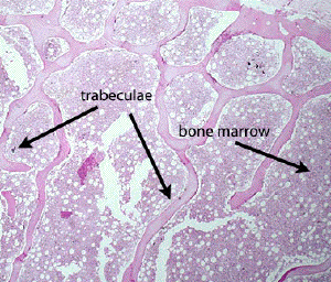
Using low power, identify: trabeculae and hematopoietic cells of the marrow. Assess the cellularity of the marrow, i.e. the proportion of hematopoietic cells compared to fatty tissue.
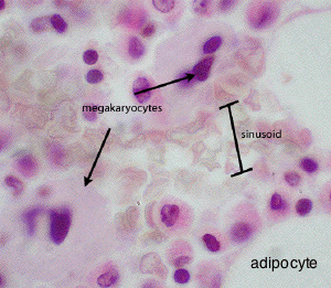
Using high power, identify in the marrow:
Venous sinusoids which are identified by the aggregations of mature RBCs in their lumens. You may also be able to see some arterioles.
Megakaryocytes. Giant cells with single large polyploid nuclei What do these cells produce?
Erythroblastic (erythroid) islands. With long, careful study of marrow specimens, an architectural organization to the seemingly random arrangement of myeloid tissue may be appreciated. Clusters of developing red blood cells, the erythroid islands, can be found in proximity to the marrow sinusoids. A macrophage is typically found in the midst of the cluster, but may be difficult to distinguish. In our slide collection, erythroid islands are best observed in the punch biopsy specimen. It may be useful to first spend time familiarizing yourself with the cells of the erythroid lineage by examining the bone marrow smear, and then return to this slide after you can more easily recognize their morphology.
The remainder of the cells consist of the granulocytic, monocytic and lymphocytic lines. You can easily see some juvenile and mature neutrophils and eosinophils. Note: this section is mainly to demonstrate general architecture of active marrow, not the detailed structure of hematopoietic cells.