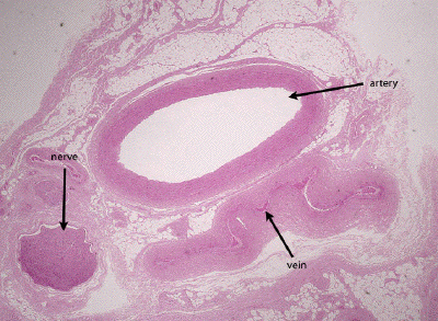
Slide #DMS 107 [Neurovascular bundle, Monkey, PTS]. This slide contains a complete neurovascular bundle (artery, vein and nerve). First, examine the medium-size, distributing ("muscular") artery. In general, the muscular arteries correspond to the smaller named arteries which you dissected in gross anatomy lab. Contrast in detail the structure of the media of these arteries to that of the aorta. Note the predominance of smooth muscle, the sparseness of elastic fibers and the prominent internal elastic lamina. What is the significance of the difference in relative amounts and organization of elastic fibers, collagenous fibers, and smooth muscle cells in terms of the functional role played by the two types of arteries in controlling circulation?
Note that the adentitia of these muscular arteries contains small blood vessels (vasa vasorum) and elastic and collagen fibers.
Next, try to identify veins. Study the relative thickness of the tunics in the walls of these veins and compare their characteristics with those of the comparable muscular artery as observed above. Next compare the distribution of elastic fibers. As compared with the accompanying artery, how do you relate the larger caliber and thinner wall of the vein to its function? Veins vary greatly in their histology according to the amount of hydrostatic pressure exerted on the wall. Thus, those located above the heart have a thinner wall than those located below the heart.
This is a very low power view of a neurovascular bundle stained with H&E. A muscular artery, accompanying vein (collapsed in this preparation) and nerve can all be seen.
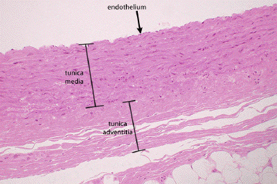
This is a low power view through the wall of a muscular artery, stained with H&E. Although elastic fibers are present in this vessel, they are not readily visualized without a specific stain for elastic tissue. The nuclei of the endothelium may be seen, and one may roughly define the tunica media and the tunica adventitia, the latter blending with the perivascular connective tissue.
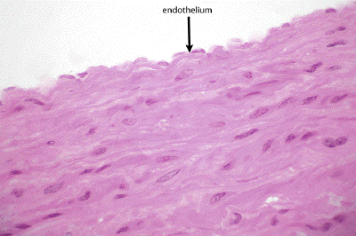
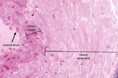
At medium power, the endothelium of this muscular artery is apparent, though the border between the tunica intima and the tunica media is not well-defined in the absence of a specific stain for the internal elastic lamina. In the media, one may find smooth muscle cells intermixed with occasional elastic fibers (not well-visualized in H&E-stained sections).
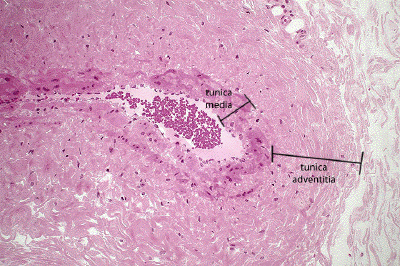
A medium power view of a large vein shows the relatively thin tunica media, comprised of just a few layers of smooth muscle, and an extensive tunica adventitia.
At high power, one can see the endothelium of the tunica intima of this large vein, the nuclei of the smooth muscle cells comprising the rather thin tunica media, and the abundant collagenous tissue forming the extensive tunica adventitia.