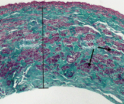
Slide #DMS 110B [vena cava, Masson trichrome]. Large veins, such as the inferior vena cava, are easily distinguished by their size, thinness of the media and the striking accumulation of longitudinally disposed smooth muscle in the adventitia.

This Masson-stained section through a vena cava demonstrates nicely the extent of the tunica adventitia (connective tissue stained green), and the bundles of longitudinally-oriented smooth muscle (stained reddish-purple) found there.