Slide #DMS 162 [Parathyroid, human, H&E]. The amount of adipose tissue in the human parathyroid is variable. The section seen here contains a good deal of adipose tissue, but the parenchymal cells, both chief cells and oxyphil cells, are well preserved and easily distinguished.
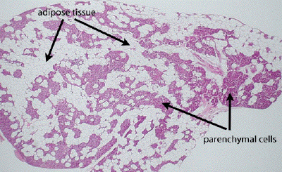
This is a very low power view through a section of human parathyroid gland. Islands of parenchymal cells are found amidst a fair amount of adipose tissue. The adipose tissue content of parathyroids can vary significantly among individuals, and does not necessarily reflect age-related fatty involution such as one sees in the thymus.
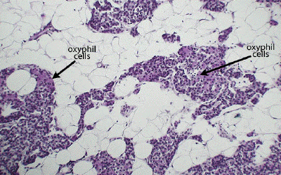
At higher power, one can easily define the adipocytes filling the spaces between clusters of parenchymal cells. Even at this power, one can distinguish the small, closely packed nuclei of the chief cells and the clusters of the large, eosinophilic oxyphil cells, seen to better effect in the next image.
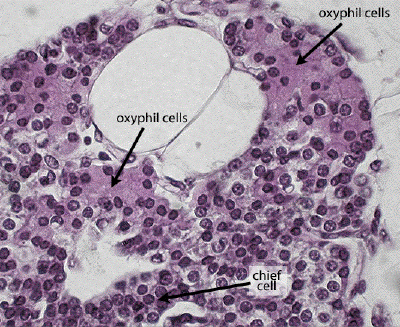
At high power, note the large clusters of large, eosinophilic oxyphil cells. Their eosinophilia is derived from a cytoplasm replete with mitochondria. Lightly stained chief cells, responsible for the production of parathyroid hormone, dominate the rest of the parenchymal tissue.