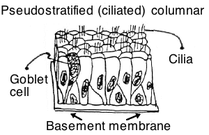
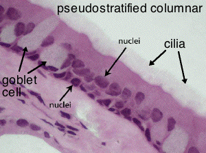
Slide DMS029. [trachea, monkey; x-section, PTS]. The lumen of the trachea is lined by pseudostratified columnar epithelium. Note that all the surface cells have cilia at their free (apical) surface. (What function do these cilia serve in the trachea?) Mucus-secreting unicellular gland cells ("goblet cells") are interspersed among the ciliated cells.
The term "pseudostratified" is commonly applied to certain epithelia which, when sectioned, appear to be stratified. However, observation of cells separated by teasing shows that all of the cells are attached to the underlying basement membrane, but only some reach the free surface of the epithelium. This kind of epithelium is most commonly found lining the passages of the respiratory system (e.g., trachea and bronchi).
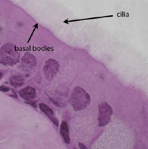
There are motile apical surface specializations called cillia on the pseudostratified columnar epithelium in the trachea (and in other places such as the oviduct). Scan the slide first at low power. Locate well-stained individual cells or clumps of cells with tufts of cilia. Observe ciliary basal bodies. Review the ultrastructure of cilia in slides #3 and #4 of the ultrastructure module of this lesson.


This oil emersion view shows the cilia and the appearance of basal bodies as a darkly staining line at the cell apex.
