Slide #DMS 174 [Ovarian follicles. monkey, H&E]. The slide of monkey ovary shows a variety of follicles in the cortex, but has no recent corpora lutea. You may see an intramural portion of the oviduct at one end of this section.
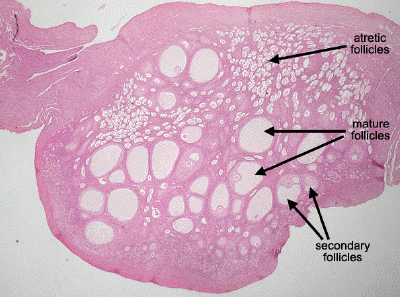
This is a very low power view through another specimen of monkey ovary. Numerous secondary follicles and mature (Graafian) follicles can be seen in this image, as well as an abundance of small atretic follicles.
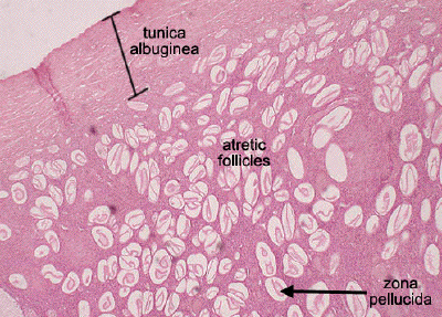
A higher power view of the outer ovarian cortex shows the tunica albuginea, beneath which is the ovarian stroma surrounding numerous atretic follicles. In these degenerating follicles, probably former primary follicles, the oocyte has disintegrated, leaving only a collapsed zona pellucida.
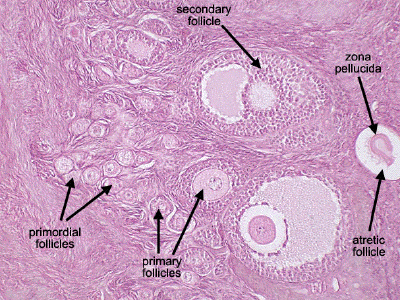
In this view of a different part of the ovarian cortex, one may identify follicles at several different stages of development including the small primordial follicles, unilaminar and multilaminar primary follicles, secondary follicles, as well as an atretic follicle containing just the remnants of the zona pellucida.
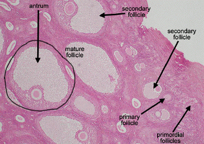
In this low power view of the ovarian cortex, one may appreciate the remarkable growth in size of the follicles as they progress from small primordial and primary follicles to secondary follicles and finally mature, Graafian follicles which exhibit a single, large antral space filled with liquor folliculi.