Slide DMS182 [Placenta, human, PTS]. This is a section of placenta in the second trimester of pregnancy. It includes the chorionic plate and attached placental villi, but no maternal tissue except for some residual blood in the intervillous spaces. (Refer to your texts and atlases for orientation).
The chorionic plate proper consists of rather dense vascular and connective tissue. In places on the fetal (nonvillous) side of the plate there are amniotic cells which are hard to see since thay have been allowed to dry before they were fixed and thus stain poorly. The maternal (villous) side of the chorionic plate gives origin to the placental villi. These are seen in various sizes and in various stages of preservation. Note the reddish band of inflammatory material at the inner surface of plate consisting mostly of neutrophils and precipitated fibrin. This is pathologic, "chorioamnionitis", probably due to an ascending infection of the birth canal. (And it was probably why this pregnancy was terminated early on).
Study the villi at higher magnification. They are covered by a continuous layer of syncytiotrophoblast. In many places tips of villi have been cut across so that the section contains islands or buds of syncytiotrophoblast. In other places the villi can be seen in cross, longitudinal, or oblique section. Fetal blood vessels are demonstrable in most villi. Surrounding the blood vessels is irregular loose connective tissue. In many of the villi cytotrophoblast cells can be seen just beneath the syncytiotrophoblast. These occur singly or in small nests. These cells divide to renew the syncytiotrophoblast. You should recall that in earlier pregnancy villi are covered by a double layer: an outer continuous syncytiotrophoblast layer and beneath that, a continuous cytotrophoblast layer. How do the two differ morphologically and functionally? Later in pregnancy the cytotrophoblast layer is almost completely lost; only a few scattered cells or nests of cells remain in the term placenta. The CT within the villi contains a population of macrophages called Hofbauer cells. In addition to their customary role as part of the immune system, these cells may help to maintain villous homeostasis by affecting water regulation and nutrient/waste transport.
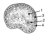
XS of placental villus, early pregnancy: 1. CT stroma with fetal blood vessels (note nucleated RBCs); 2. Hofbauer cell; 3. Cytotrophoblast; 4. Syncytiotrophoblast