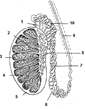
Slide DMS186 [Testis, human, H&E or Masson's]. Both of these slides are good for studying the general structure of the organ. Identify the following: tunica albuginea, seminiferous tubules, interstitial CT and interstitial (Leydig) cells. Reserve detailed study of the seminiferous epithelium for the next slide. In the H&E slide you will see the mediastinum testis with its epithelium-lined channels, the rete testis, into which the seminiferous tubules lead. The Masson-stained section provides a good view of the interstitial CT, blood vessels, and interstitial (Leydig) cells. In the latter, look for the crystals of Reinke which are peculiar to humans but of unknown significance. What hormone is produced by the Leydig cells and what hormone stimulates its synthesis and secretion?

Longitudinal section of testis, Epididymis, ductus deferens: 1. Ductuli efferentes; 2. Tubulus rectus; 3. Seminiferous tubule; 4. Interlobular septum; 5. Tunica albuginea; 6. Caudal epididymis; 7. Corpus epidiymis; 8. Mediastinum, rete testis; 9. Ductus deferens; 10. Caput epididymis