Slide DMS151 [Monkey; Kidney]. This full-thickness cross section of the kidney of a small monkey serves well as an orientation slide. Although the monkey has a multilobar kidney like humans, when cut in cross section it resembles a unilobar kidney and is thus relatively easy to interpret. First Identify capsule, cortex, medulla, and hilum regions. Within the cortex define medullary rays and cortical labyrinth. Observe that the medullary rays consist chiefly of radially oriented tubules, whereas the labyrinth contains both convoluted tubules and globose renal corpuscles. Each medullary ray forms the central axis of a renal lobule but does not extend all the way to the capsule.
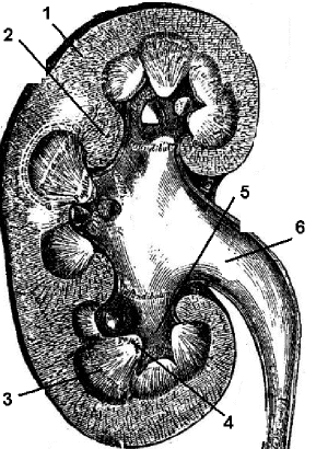
The medulla is subdivided into renal pyramids, each along with overlying cortex the basic unit of a renal lobe. Again, only one lobe and one pyramid is seen in this particular section. The base of the pyramid is contiguous with the cortex, the apex is oriented toward the pelvis and projects into a minor calyx. Note how the cortex extends down around the sides of the pyramid. In longitudinal sections these cortical extensions between pyramids are denoted as the renal columns (of Bertin).
In the hilar region, note how the minor calyx cups around the apex (papilla) of a pyramid. You may see papillary ducts (of Bellini) opening into the calyx at the papillary apex (area cribrosa). Examine the histology of the wall of calyx. What is the functional significance of the transitional epithelium (poorly preserved in this slide) and underlying smooth muscle? Observe the white adipose tissue that fills the surrounding pelvic sinus. Identify the renal artery, vein and associated nerves.
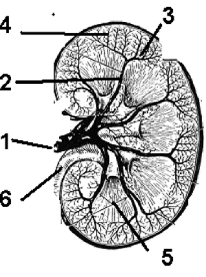
Referring to the drawings in the lab guide, trace the parts of the renal vascular system. Now, examine your slide and look for the following vessels: (a) interlobar, between adjacent pyramids, (b) arcuate, at corticomedullary boundary, (c) interlobular, midway between neighboring medullary rays, (d) afferent and efferent arterioles of the renal corpuscles, which usually cannot be differentiated from each other with ease, and (e) the capillary plexuses which surround tubules in the cortex and medulla and are fed respectively by the efferent glomerular arteriole and the vasa recta (arteriolae rectae [spuriae ]). The arteries are accompanied by corresponding veins.
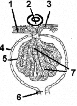
Renal Corpuscle. The renal corpuscles are well-represented in this slide, though the subsequent tubular elements of the nephron will be best examined in subsequent slides. Locate in the cortical labyrinth a renal corpuscle which consists of a glomerulus and a capsule(of Bowman). Identify the visceral and the parietal epithelial layers of the glomerular capsule.

Renal corpuscle: 1. afferent arteriole; 2. macula dense of distal tubule; 3. efferent arteriole; 4. Bowman?fs capsule (parietal layer); 5. visceral layer ?| podocytes; 6. proximal tubule; 7. glomerulus cappillaries
Study in detail the relationship of the visceral layer of the capsule to the endothelium of the peculiar arterial capillaries which constitute the glomerulus. Understand the structure of this filtration barrier as revealed by electron microscopy (See illustrations below and illustrations in your textbook). Where are mesangial cells located and what functions do they serve?
Identify the vascular pole of the renal corpuscle in relation to the afferent and efferent arterioles of the glomerulus. Look for a macula densa where a segment of distal convoluted tubule abuts the vascular pole. Where are the other components of the juxtaglomerular apparatus (JG cells, extraglomerular mesangium) located? (Note: demonstration of JG cell granules requires special staining techniques). What is the function of the JG apparatus?
Find a corpuscle that shows the point of continuity between the parietal epithelium of the glomerular capsule and the epithelium of a proximal convoluted tubule , ie., the urinary pole, noting the change in epithelial morphology.
Referring to the drawing below, trace the parts of the renal vascular system. Now, examine your slide and look for the following vessels: (a) interlobar, between adjacent pyramids, (b) arcuate, at corticomedullary boundary, (c) interlobular, midway between neighboring medullary rays, (d) afferent and efferent arterioles of the renal corpuscles, which usually cannot be differentiated from each other with ease, and (e) the capillary plexuses which surround tubules in the cortex and medulla and are fed respectively by the efferent glomerular arteriole and the vasa recta (arteriolae rectae [spuriae ]). The arteries are accompanied by corresponding veins.

Arterial Supply to the Kidney: 1. Renal artery [leading to segmental arteries]; 2. Interlobar artery [within the renal column; 3. Arcuate artery [at the cortico-medullary jx]; 4. Intralobular artery [leading to glomerulus]; 5. Renal pyramid [medulla]; 6. ureter [formed from renal pelvis]

Human Kidney: 1. Cortex; 2. Renal columns; 3. Medulla; 4. Papilla of medulla; 5. Calyx; 6. Renal pelvis.