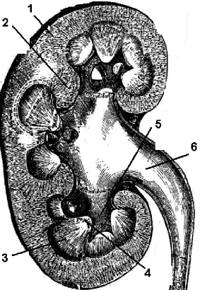
Slide DMS152 [Rabbit; Kidney, pelvis and ureter, l.s., H&E]. Orient yourself to this slide by identifying the features with which you are already familiar. This slide shows a partial longitudinal section of a unilobar kidney. It is a relatively thick section but shows some details of kidney structure very well. Hold the slide up to light and and view with your unaided eye or with an inverted ocular. You can readily identify cortex, and the various zones of the medulla: outer medulla (further divided into outer and inner stripes), inner medulla, papilla, and then calyx and ureter. Switch to the high power and focus on the papilla, calyx, and renal sinus regions. The transitional epithelium (urothelium) covering the papilla and lining the calyx is well preserved in this specimen. Look for segments of the ureter that drain the calyces. What type of lining epithelium do they present?
Now return to your study of the uriniferous tubule. First, identify the renal corpuslces, which, in this preparation, are not so elegantly dislpayed as in Slide #DMS151. Now turn your attention to the tubular system of the cortex. Although better preservation of tubular architecture will be seen in subsequent slides, some of the fundamentals may be appreciated here. Identify the proximal convoluted tubules, the profiles of which dominate the cortical labyrinth. Study the epithelium for the following characteristics: size and shape of cells, density of cytoplasmic granulation and presence of a brush border. (The brush border is very sensitive to fixation conditions and was not well-preserved in this section). How many nuclei are usually found in a cross-section of the tubule? In a medullary ray identify the straight part of the proximal tubule (aka, thick descending limb of loop of Henle) and note that the cytology is the same as that of the convoluted portion.
In the outer zone of the medulla identify the thin limbs of the loop of Henle and contrast the size of the tubule and thickness of its epithelium with those of neighboring capillaries. (The capillaries usually contain some RBCs). The thin portion of Henle's loop is especially clear in the inner zone of the medulla.
In the outer medullary zone and in a medullary ray, try to find the thick part of an ascending thick limb of Henle's loop (aka: straight part of distal tubule). Compare its histology with that of the proximal segment of the tubule. These differences can be discerned quickly in the medullary ray because the distal and proximal segments are the only parts of the uriniferous tubule present whose epithelial cells have a granular cytoplasm.
Review how the arrangement of the different segments of the loop of Henle in relation to vasa recta establishes a "countercurrent exchange system" for adjusting the osmotic concentration of the blood in the medulla.
Study the distal convoluted tubule in the cortical labyrinth. Except for the convoluted form, its structure is identical with that of the thick ascending portion of Henle's loop in the medullary ray. Again, the preservation of cellular architecture is not optimal in this section, and some of these features will be more apparent in subsequent slides.
Collecting System. Within medullary rays find cortical collecting tubules. In comparison with the proximal and distal convoluted tubule, note the caliber of the lumen, uniformity in height of the epithelium and size of its cells, cytoplasmic homogeneity, and distinctness of the cell membranes. The major changes seen as they continue into the medullary collecting tubule s and then the papillary ducts are a gradual enlargement of the lumen and an increase in the height of the lining epithelium. Locate an opening of papillary duct into a minor calyx at the area cribrosa.
Observe the character of the interstitial connective tissue in relation to tubules and blood vessels. In which part of the kidney is connective tissue most abundant?

Human Kidney: 1. Cortex; 2. Renal columns; 3. Medulla; 4. Papilla of medulla; 5. Calyx; 6. Renal pelvis.