Slide DMS153 [Kidney, human, x. s., Masson's or H&E]. The Masson's-stained section is more instructive than the H&E so be certain to see it. First examine the slide with low and high power. Note that your section contains only a small portion of the cortex and medulla. The Masson-stained preparation demonstrates well the relative abundance of connective tissue in cortex and medullas (collagen stains green). Switch to your 40x objective and study the cortical region. Components of nephrons are well preserved. Proximal and distal convoluted tubules are readily distinguished. Note the luxuriant brush border and deeply stained epithelial cells of the proximal tubule. Details of renal corpuscle structure are easily seen. Now study the medulla. Note how the vasa rectae tend to run in bundles surrounded by much connective tissue. Collecting ducts, and thin limbs of Henle's loop can be easily identified. Note the distinct cell boundaries of the epithelial cells in the former.

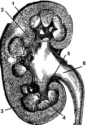
Human Kidney: 1. Cortex; 2. Renal columns; 3. Medulla; 4. Papilla of medulla; 5. Calyx; 6. Renal pelvis.