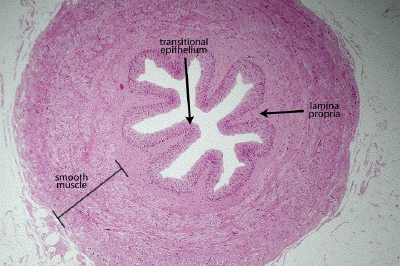
Slide #DMS 154 [Ureter, Human, H&E]. This slide shows the characteristic transitional epithelium very well. Note that some of the cells are binucleate or have an unusually large nucleus. Terminal bars are distinct. Although this epithelium resembles stratified squamous epithelium there are many important structural and functional differences. In the undistended ureter the mucosa tends to thown into folds. Note that the ureter has a lamina propria, but no submucosa The muscularis (all smooth muscle) is extensive and interlaced with connective tissue and the orientation of the muscle bundles is rather haphazard. In general, there tends to be an inner longitudinal and an outer circular layer. Why is there such an abundance of smooth muscle in the ureter? Does the ureter have a serosal or adventitial outermost layer? In cross section the lumen is typically stellate in shape. Looking at the shape of the surface cells of the transitional epithelium , would you judge that the ureter was fixed in a contracted or distended state? Does this section have 2 (or 3?) layers of muscle in the muscularis externa?
This is a very low power section through a monkey ureter. Note the lumen lined by transitional epithelium, beneath which is a layer of lamina propria and then a rather thick wall replete with smooth muscle disposed in a variety of ill-defined orientations, though with a tendency towards an inner longitudinal and outer circular layer (opposite to the orientation seen in the gut).
.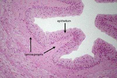
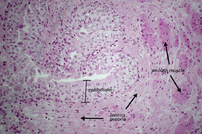
At higher power, one can appreciate the stratified appearance of the transitional epithelium, including the somewhat odd shapes assumed by the apical-most layer of cells in the epithelium. Supporting the epithelium is the lamina propria consisting of a rather dense fibroelastic connective tissue network.
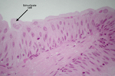
A high power view of the ureteral lumen shows the architecture of the transitional epithelium. Note the odd, sometimes angular or 'umbrella' like shape typical of the apical-most layer of cells in a non-distended viscus. Binucleate cells are not uncommon
.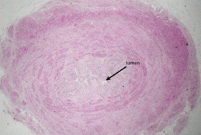
This is a very low power view of a section through the human ureter, the lumen of which is largely collapsed.
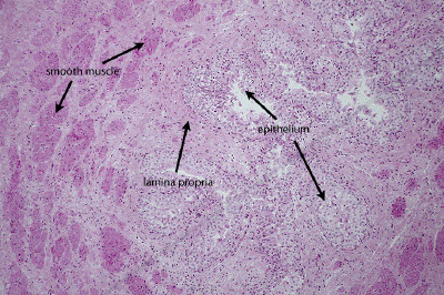
At low power, one can discern the transitional epithelium lining the somewhat collapsed lumen of the ureter. A rather dense, fibroelastic lamina propria supports the epithelium. Some bundles of generally longitudinally-oriented smooth muscle can be seen in the inner portion of the wall of this organ.
At higher power, appreciate transitional epithelium and note the variety of cell shapes assumed by the apical-most layer of cells. The dense lamina propria and more eosinophilic bundles of smooth muscle are also apparent.
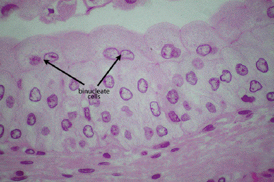
At high power, appreciate the classic morphology of the apical-most cells of the transitional epithelium from an organ in a relaxed (non-distended) state. In addition to a pillow or umbrella-like morphology, many cells may be binucleate.